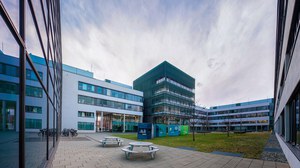
Jochen Guck - Cellular Machines

Cells are the basic entities of biological systems. They can be seen as little machines that have particular physical properties, which enable them to navigate their 3D physical environment and fulfil their biological functions. We investigate these physical - mechanical and optical - properties of living cells and tissues using novel photonics and biophysical tools to test their biological importance. Our ultimate goal is the transfer of our findings to medical application in the fields of improved diagnosis of diseases and novel approaches in regenerative medicine.
Research
While most current biological research focuses on molecular and biochemical aspects of cells and their functioning, we are interested in their global physical properties and how the cells relate to the physical properties of their environment.
Cell mechanics
Our recent findings increasingly demonstrate that the mechanical properties of cells determine the physical limits of cell function - for example in 3D cell migration. Cell mechanics can thus be used to characterise cells, to monitor physiological changes (such as stem cell differentiation), and to diagnose pathological alterations (such as metastatic progression or inflammatory reactions). Publication: F. Lautenschläger et al., The regulatory role of cell mechanics for migration of differentiating myeloid cells. Proc. Natl. Acad. Sci. U.S.A. 106:15696-15701 (2009); T.W. Remmerbach et al., Oral Cancer Diagnosis by Mechanical Phenotyping. Cancer Res. 69:1728–1732 (2009).
The optical stretcher is a novel laser tool that can be used to trap and deform individual biological cells in order to test such mechanical properties. Ultimately, we are developing a label-free, high-throughput cell analysis method for cancer and sepsis diagnosis and for stem cell sorting.

Schematic of an optical stretcher. In a microfluidic flow chamber, cells in suspension (green) can be trapped by two opposing laser beams (red) of low intensity, emanating from optical fibers (blue). Increasing the intensity of the laser light augments the forces at the surface of the cell, leading to measurable deformation. Publication: J. Guck et al., Optical deformability as an inherent cell marker for testing malignant transformation and metastatic competence Biophys. J. 88:3689-3698 (2005)
Mechanosensing and Tissue Mechanics in the CNS
It is increasingly recognised that cells react to the mechanical properties of their environment and that these mechanical cues can be as important as adhesive or soluble biochemical cues. We are especially interested in assessing the importance of this "mechanosensing" in the development and in pathological conditions in the central nervous system. For a recent review, please see: K. Franze and J.Guck, The biophysics of neuronal regrowth Rep. Progr. Phys. 73:094601 (2010)
In order to characterise and quantify the mechanical environment of cells in the central nervous system with high spatial resolution, we have developed the mechanical mapping of CNS tissue using scanning force microscopy. Publication: A. Christ et al., Mechanical difference between white and grey matter in the rat cerebellum measured by scanning force microscopy J. Biomechanics 43:2986-2992 (2010)
Retina Optics
Another example for the importance of physics in biology are the optical properties of cells, specifically in the retina, which is, curiously, inverted with respect to its optical function. The light-sensing photoreceptor cells are located on the 'wrong' side - the side furthest away from the incoming light. Consequently, light has to traverse hundreds of microns of potentially scattering tissue. We have shown that there are cells in the retina that act as optical fibers and that photoreceptor cells even invert their usual nuclear chromatin arrangement to turn them into microlenses. Both aspects improve the light transmission through the retina and shed new light on the retina as an optical system. Publications: K. Franze et al., Müller cells are living optical fibers in the vertebrate retina. Proc. Natl. Acad. Sci. U.S.A. 104:8287-9292 (2007); I. Solovei et al., Nuclear architecture of rod photoreceptor cells adapts to vision in mammalian evolution. Cell 137:356-368 (2009).
Publications
1. K. J. Chalut, A. E. Ekpenyong, W. L. Clegg, I. C. Melhuish, and J. Guck. Quantifying cellular differentiation by physical phenotype using digital holographic microscopy. Integr. Biol., DOI: 10.1039/C2IB00129B (2012).
2. J. da Silva, F. Lautenschläger, C.-H. R. Kuo, J. Guck and E. Sivaniah. 3D inverted colloidal crystals in realistic cell migration assays for drug screening applications. Integr. Biol. 3(12):1202-1206 (2011).
3. L. Boyde, K.J. Chalut, and J. Guck. Exact analytical expansion of an off-axis Gaussian laser beam using the translation theorems for the vector spherical harmonics. Appl. Opt. 50(7):1023-1033 (2011).
4. L. Boyde, K.J. Chalut, and J. Guck. Near- and far-field scattering from arbitrary three-dimensional aggregates of coated spheres using parallel computing. Phys. Rev. E 83:026701 (2011).
5. K. Franze, M. Francke, K. Günter, A.F. Christ, N. Körber, A. Reichenbach, and J. Guck. Spatial mapping of the mechanical properties of the living retina using scanning force microscopy. Soft Matter, 7(7):3147–3154 (2011).
6. J. Guck. Cell Sorting, in: Handbook of Biophotonics, Vol. 2: Photonics for Health Care, ed. by J. Popp, V.V. Tuchin, A. Chiou, and S. Heinemann, Wiley-VCH, Berlin (2011).
7. J.M. Maloney, D. Nikova, F. Lautenschläger, E. Clarke, R. Langer, J. Guck, and K.J. Van Vliet. Mesenchymal stem cell mechanics from the attached to the suspended state. Biophys. J. 99(8):2479-2487 (2010).
8. K. Franze and J. Guck. The biophysics of neuronal regrowth. Rep. Prog. Phys. 73:094601 (2010).
9. J. Guck, F. Lautenschläger, S. Paschke, and M. Beil. Critical review: Cellular mechanobiology and amoeboid migration. Integr. Biol. 2(11):575-583 (2010).
10. A.F. Christ, K. Franze, H. Gautier, P. Moshayedi, J. Fawcett, R.J.M. Franklin, R.T. Karadottir, and J. Guck. Mechanical difference between white and gray matter in the rat cerebellum measured by scanning force microscopy. J. Biomech. 43:2986–2992 (2010).
11. M. Kreysing, L. Boyde, J. Guck, and K. Chalut. Physical insight into light scattering by photoreceptor cell nuclei. Opt. Lett. 35(15):2639-2641 (2010).
12. J.M.A Mauritz, A. Esposito, T. Tiffert, J.N. Skepper, A. Warley, Y.Z. Yoon, P. Cicuta, R., V.L. Lew, J. Guck, and C.F. Kaminski. Biophotonic techniques for the study of malaria-infected red blood cells. Med. Biol. Engin. Comp. 48(10): 1055-1063 (2010).
13. J.M.A Mauritz, T. Tiffert, R. Seear, F. Lautenschläger, A. Esposito, V.L. Lew, J. Guck, and C.F. Kaminski. Detection of Plasmodium falciparum-infected red blood cells by optical stretching. J. Biomed. Opt. 15:030517 (2010).
14. B. Kemper, P. Langehanenberg, A. Höink, G. von Bally, F. Wottowah, S. Schinkinger, J. Guck, J. Käs, I. Bredebusch, J. Schnekenburger, and K. Schütze. Monitoring of Laser Micromanipulated Optically Trapped Cells by Digital Holographic Microscopy.
15. P. Moshayedi, L. da F Costa, A. Christ, S. P. Lacour, J. Fawcett, J. Guck, and K. Franze. Mechanosensitivity of astrocytes on optimized polyacrylamide gels analyzed by quantitative morphometry. J. Phys.: Cond. Matter 22(19):194114 (2010).
16. J. da Silva, F. Lautenschläger, E. Sivaniah, and J. Guck. The cavity-to-cavity migration of leukaemic cells through 3D honey-combed hydrogels with adjustable internal dimension and stiffness. Biomaterials 31(8):2201–2208 (2010).
17. I. Solovei, M. Kreysing, Ch. Lanctôt, S. Kösem, L. Peichl, Th. Cremer, J. Guck*, and B. Joffe*. Nuclear architecture of rod photoreceptor cells adapts to vision in mammalian evolution. Cell 137(2):356-68 (2009) [* joint corresponding authors].
18. J. Guck. Do cells care about physics? Physics World 22(7): 31-34 (2009).
19. F. Lautenschläger, S. Paschke, A. Bruel, S. Schinkinger, M. Beil, and J. Guck. The regulatory role of cell mechanics for migration of differentiating myeloid cells. Proc. Natl. Acad. Sci. U.S.A. 106(37):15696-15701 (2009).
20. L. Boyde, K. Chalut, and J. Guck. Interaction of Gaussian Beam with Near-Spherical Particle: an Analytic-Numerical Approach for Assessing Scattering and Stresses. JOSA A 26(8):1814–26 (2009).
21. T.W. Remmerbach, F. Wottawah, J. Dietrich, B. Lincoln, Ch. Wittekind, and J. Guck. Oral Cancer Diagnosis by Mechanical Phenotyping. Cancer Res. 69(5):1728–1732 (2009).
22. M. K. Kreysing, T. Kießling, A. Fritsch, Ch. Dietrich, J. R. Guck, J. A. Käs. The optical cell rotator. Opt. Express 16(21): 16984-16992 (2008).
23. K. Franze, J. Grosche, S. N. Skatchkov, S. Schinkinger, D. Schild, O. Uckermann, K. Travis, A. Reichenbach, and J. Guck. Müller Cells are Living Optical Fibers in the Vertebrate Retina. Proc. Natl. Acad. Sci. U.S.A. 104(20):8287-8292 (2007).
24. B. Lincoln, S. Schinkinger, F. Wottawah, and J. Guck. High-Throughput Cell Analysis with a Microfluidic Optical Stretcher. Meth. Cell Biol. 83:397-423 (2007).
25. B. Lincoln, S. Schinkinger, K. Travis, F. Wottawah, S. Ebert, F. Sauer, and J. Guck. Reconfigurable microfluidic integration of a dual-beam laser trap with biomedical applications. Biomed. Microdev. 9(5):703-710 (2007).
26. S. Ebert, K. Travis, B. Lincoln, J. Guck. Fluorescence Thermometry in a Microfluidic Dual-Beam Laser Trap. Opt. Express 15(23):15493-15499 (2007).
27. R. Ananthakrishnan, J. Guck, F. Wottawah, S. Schinkinger, B. Lincoln, M. Romeyke, T. Moon, and J. Käs. Quantifying the Contribution of Actin Networks to the Elastic Strength of Fibroblasts. J. Theo. Biol. 242:502–516 (2006).
28. Y. Lu, K. Franze, G. Seifert, Ch. Steinhäuser, F. Kirchhoff, H. Wolburg, J. Guck, P. Janmey, E. Wei, J. Käs, and A. Reichenbach. Viscoelastic properties of individual glial cells and neurons in the CNS. Proc. Natl. Acad. Sci. U.S.A. 103(47):17759–177764 (2006).
29. R. Ananthakrishnan, J. Guck, and J. Käs. Cell mechanics: Recent advances with a theoretical perspective, in: Recent Research Developments in Biophysics, Transworld Research Network 5:39-69 (2006)
30. K. Travis and J. Guck. Scattering from Single Nanoparticles: Mie theory revisited. Biophys. Rev. Lett. 1(2):115–143 (2006).
31. F.-U. Gast, P. S. Dittrich, P. Schwille, M. Weigel, M. Mertig, J. Opitz, U. Queitsch, S. Diez, B. Lincoln, F. Wottawah, S. Schinkinger, J. Guck, J. Käs, J. Smolinski, K. Salchert, C. Werner, C. Duschl, M. S. Jäger, K. Uhlig, P. Geggier, S. Howitz. The microscopy cell (MicCellTM), a versatile modular flowthrough system for cell biology, biomaterial research, and nanotechnology. Microfluid. Nanofluid. 2(1):21–36 (2006).
32. F. Wottawah, S. Schinkinger, B. Lincoln, R. Ananthakrishnan, M. Romeyke, J. Guck, and J. Käs. Optical Rheology of Biological Cells. Phys. Rev. Lett. 94(9):98103 (2005)
33. F. Wottawah, S. Schinkinger, B. Lincoln, S. Ebert, K. Müller, F. Sauer, K. Travis, and J. Guck. Characterizing Single Suspended Cells by Optorheology. Acta Biomat. 1:263–271 (2005).
34. J. Guck, H. Erickson, R. Ananthakrishnan, D. Mitchell, M. Romeyke, S. Schinkinger, F. Wottawah, B. Lincoln, J. Käs, S. Ulvick, and C. Bilby. Optical Deformability as Inherent Cell Marker for Testing Malignant Transformation and Metastatic Competence. Biophys. J. 88(5):3689-3698 (2005).
35. R. Ananthakrishnan, J. Guck, F. Wottawah, S. Schinkinger, B. Lincoln, M. Romeyke, T.J. Moon, and J Käs. Modelling the structural response of an eukaryotic cell in the optical stretcher. Curr. Sci. 88(9):1434–1440 (2005).
36. T. Betz, J. Teipel, D. Koch, J. Guck, J. Käs, H. Gießen. Excitation beyond the monochromatic laser limit: Simultaneous 3-D confocal and multiphoton microscopy with a tapered fiber as white-light laser source. J. Biomed. Opt. 10(5):054009 (2005)
37. B. Lincoln, H. M.Erickson, S. Schinkinger, F. Wottawah, D. Mitchell, S. Ulvick, C. Bilby, and J. Guck. Deformability-Based Flow Cytometry. Cytometry A 59:203-209 (2004).
38. S. Schinkinger, K.A. Travis, F. Wottawah, B. Lincoln, and J. Guck. Feeling for Cells with Light. Proc. SPIE: Optical Trapping and Optical Micromanipulation 5514:170-178 (2004).
39. J. Guck, R. Ananthakrishnan, C.C. Cunningham, and J. Käs. Stretching Biological Cells with Light. J. Phys. Cond. Mat. 14: 4843-4856 (2002).
40. J. Guck, R. Ananthakrishnan, H. Mahmood, T.J. Moon, C.C. Cunningham, and J. Käs. The Optical Stretcher: A Novel Laser Tool to Micromanipulate Cells. Biophys. J. 81(2):767–784 (2001).
41. J. Guck. Optical Deformability–Micromechanics from Cell Research to Biomedicine. Ph.D. dissertation University of Texas at Austin, U.S.A. (2001).
42. J. Guck, R. Ananthakrishnan, T.J. Moon, C.C. Cunningham, and J. Käs. Optical Deformability of Soft Biological Dielectrics. Phys. Rev. Lett. 84(23):5451-5454 (2000)
43. J. Käs, J. Guck, and D. Humphrey, Dynamics of Single Protein Polymers Visualized by Fluorescence Microscopy, in: Modern Optics, Electronics and High Precision Techniques in Cell Biology, ed. by G. Isenberg, Springer Verlag, 103-137 (1997).
Curriculum Vitae
- 2001 PhD in Physics, University of Texas at Austin
- 2002 Junior group leader, Universität Leipzig
- 2007 Lecturer, Cavendish Laboratory, University of Cambridge
- 2009 Reader, Cavendish Laboratory, University of Cambridge
- 2012 Principal research scientist, University of Cambridge
- 2012 Professor of Cellular Machines, Biotechnology Center, TU-Dresden
- 2012 Alexander von Humboldt Professor
Positions
I am currently looking for PhD students and postdocs in the fields of biophotonics and tissue mechanics. Applicants with backgrounds in physics, bioengineering, cell or molecular biology, and medicine are welcome.
Applicants for ERC Starting Grants and DFG Emmy Noether Fellowships in relevant scientific areas are encouraged to contact me to discuss ideas, and for grant writing support, lab space and leveraging funds.
Available is also a position as a technician in an ERC funded project for up to 5 years. Proven expertise in optics, electronics and/or programming is required.
Frontcovers


Group Members
You can find a list of current group members here.
Physics-Proseminar "Physics aspects in biology"
- SS 2012 (4. Semester)
- Location: Biotechnology Center, Tatzberg 47/49
- Prof. Dr. Jochen Guck/Dr. Oliver Otto
- Contact: jochen.guck(at)biotec.tu-dresden.de
- Inscription deadline: 24. February 2012
- Further information: Please send an email with a ranked list of 2-3 topics by this date to the contact email above. The topics will be assigned and communicated by return email by the end of February. In general, you should contact the tutor, who will be assigned to each talk, at least one week ahead of your date with a detailed outline of your presentation. Talks will be 30 min + 15 min scientific discussion and feedback. The language of the course is English.
Possible topics and literature suggestions:
1. Revisiting the Central Dogma One Molecule at a Time
- Bustamante, C., Cheng, W., & Meija, Y. X. (2011). Revisiting the Central Dogma One Molecule at a Time. Cell, 144(4), 480-497.
- Bustamante, C., Bryant, Z., & Smith, S. B. (2003). Ten years of tension: single-molecule DNA mechanics. Nature, 421, 423-427.
2. Direct force measurements on DNA in a solid-state nanopore
- Keyser, U. F., Koeleman, B. N., van Dorp, S., Krapf, D., Smeets, R. M. M., Lemay, S. G., Dekker, N. H., et al. (2006). Direct force measurements on DNA in a solid-state nanopore. Nature Physics, 2(7), 473-477.
- Keyser, U. F., van Dorp, S., & Lemay, S. G. (2010). Tether forces in DNA electrophoresis. Chemical Society reviews, 39(3), 939-947.
3. Single-Molecule Micromanipulation Techniques
- Neuman, K. C., Lionnet, T., & Allemand, J.-F. (2007). Single-Molecule Micromanipulation Techniques. Annual Review of Materials Research, 37(1), 33-67.
4. The role of DNA biomechanics in the regulation of gene expression.
- Milstein, J. N., & Meiners, J.-C. (2011). On the role of DNA biomechanics in the regulation of gene expression. J R Soc Interface 8:1673-1681
5. The optical stretcher: a novel laser tool to micromanipulate cells.
- Guck, J., Ananthakrishnan, R., Moon, T. J., Cunningham, C. C., & Käs, J. (2000). Optical deformability of soft biological dielectrics. Phys Rev Lett, 84(23), 5451–5454.
- Guck, J., Ananthakrishnan, R., Mahmood, H., Moon, T. J., Cunningham, C. C., & Käs, J. (2001). The optical stretcher: a novel laser tool to micromanipulate cells. Biophysical Journal, 81(2), 767-784.
6. The equilibrium shapes of phospholipid vesicles
- Seifert, U. (1997). "Configurations of fluid membranes and vesicles." Adv Fluid Membranes Vesicles 46(1): 13-137.
7. The physics of cancer
- Wirtz, D., Konstantopoulos, K., & Searson, P. C. (2011). The physics of cancer: the role of physical interactions and mechanical forces in metastasis. Nature reviews Cancer, 11, 512–522.
- Guck, J., Schinkinger, S., Lincoln, B., Wottawah, F., Ebert, S., Romeyke, M., Lenz, D., et al. (2005). Optical deformability as an inherent cell marker for testing malignant transformation and metastatic competence. Biophysical Journal, 88(5), 3689–3698.
- Remmerbach, T. W., Wottawah, F., Dietrich, J., Lincoln, B., Wittekind, C., & Guck, J. (2009). Oral cancer diagnosis by mechanical phenotyping. Cancer Research, 69(5), 1728–1732. doi:10.1158/0008-5472.CAN-08-4073
8. How cells feel and respond to the mechanical properties of their environment
- Discher, D. E., Janmey, P., & Wang, Y.-L. (2005). Tissue Cells Feel and Respond to the Stiffness of Their Substrate. Science, 310(5751), 1139–1143.
- Wozniak, M. A., & Chen, C. S. (2009). Mechanotransduction in development: a growing role for contractility. Nature Reviews Molecular Cell Biology, 10(1), 34–43.
- Marcq, P., Yoshinaga, N., & Prost, J. (2011). Rigidity Sensing Explained by Active Matter Theory. Biophysical Journal, 101(6), L33–L35.
9. Life at small Reynolds number
- Purcell, E. (1977). Life at Low Reynolds-Number. American Journal of Physics, 45(1), 3–11.
10. Bacterial chemotaxis
- Berg, H. C., E. Coli in Motion (Springer, NY, 2004) 133pp.
- Berg, H. C., Motile behaviour of bacteria. Physics Today, 53(1), 24-29.
- Alon, U., Surette, M. G., Barkai, N., & Leibler, S. (1999). Robustness in bacterial chemotaxis. Nature, 397(6715), 168–171.
11. Pattern formation in biological systems
- Koch, A. J., & Meinhardt, H. (1994). Biological pattern formation: from basic mechanisms to complex structures. Reviews of Modern Physics, 66(4), 1481–1507.
12. Optical traps in biology
- Svoboda, K. and S. M. Block (1994). "Biological applications of optical forces." Annu Rev Biophys Biomol Struct 23: 247-285.
13. Super-resolution microscopy: principles and use in biology
- Schermelleh, L., Heintzmann, R., & Leonhardt, H. (2010). A guide to super-resolution fluorescence microscopy. J Cell Biol, 190(2), 165–175.
- Huang, B., Bates, M., & Zhuang, X. (2009). Super-Resolution Fluorescence Microscopy. Annual Review of Biochemistry, 78(1), 993–1016.
14. The physics of hearing
- Bell, A. (2004). Hearing: Travelling Wave or Resonance? PLoS Biology, 2(10), e337.
- Plaçais, P. Y., Balland, M., Guérin, T., Joanny, J. F., & Martin, P. (2009). Spontaneous Oscillations of a Minimal Actomyosin System under Elastic Loading. Phys Rev Lett, 103(15).
- Camalet, S., Duke, T., Jülicher, F., & Prost, J. (2000). Auditory sensitivity provided by self-tuned critical oscillations of hair cells. Proceedings of the National Academy of Sciences of the United States of America, 97(7), 3183–3188.
15. Kinetic proofreading: why there are not more mistakes in copying DNA
- Hopfield, J. J. (1974). Kinetic proofreading: a new mechanism for reducing errors in biosynthetic processes requiring high specificity. Proceedings of the National Academy of Sciences of the United States of America, 71(10), 4135–4139.
- Yan, J., Magnasco, M. O., & Marko, J. F. (1999). A kinetic proofreading mechanism for disentanglement of DNA by topoisomerases. Nature, 401(6756), 932–935.
16. Physical limits on the size of cells – and why it pays to get together
- Short, M. B., Solari, C. A., Ganguly, S., Powers, T. R., Kessler, J. O., & Goldstein, R. E. (2006). Flows driven by flagella of multicellular organisms enhance long-range molecular transport. Proceedings of the National Academy of Sciences of the United States of America, 103(22), 8315–8319.
- Solari, C. A., Ganguly, S., Kessler, J. O., Michod, R. E., & Goldstein, R. E. (2006). Multicellularity and the functional interdependence of motility and molecular transport. Proceedings of the National Academy of Sciences of the United States of America, 103(5), 1353.
17. How does a cell find its middle?
- Tran, P., Marsh, L., Doye, V., Inoue, S., & Chang, F. (2001). A mechanism for nuclear positioning in fission yeast based on microtubule pushing. J Cell Biol, 153(2), 397–412.
- Howard, M. & Kruse, K. (2005). Cellular organization by self-organization: mechanisms and models for Min protein dynamics. J Cell Biol, 168(4), 533–536.
18. Quantum mechanical phenomena in biology
- Davies, P. (2009). The quantum of life. Physics World, July, 24-28.
- Lloyd. S. (2011). A bit of quantum hanky-panky. Physics World, January, 26-29.
19. How nerve signals propagate
- Biological Physics: Energy, Information, Life, Nelson P (WH Freeman 2003).
- Heimburg, T. (2009). Die Physik von Nerven. Physik J, 8(3), 33–39.
20. Brownian ratchets in biology
- Bier, M. (1997). Brownian ratchets in physics and biology. Contemporary Physics, 38(6), 371–379.
- Reimann, P., & Hänggi, P. (2002). Introduction to the physics of Brownian motors. Applied Physics A, 75(2), 169–178.
- Peskin, C., Odell, G., & Oster, G. (1993). Cellular motions and thermal fluctuations: the Brownian ratchet. Biophysical Journal, 65, 316–324.
- Astumian, R. D. (1997). Thermodynamics and Kinetics of a Brownian Motor. Science, 276(5314), 917–922.
General literature for the biological background:
- Essential Cell Biology, Alberts B et al. (Garland 2003).


